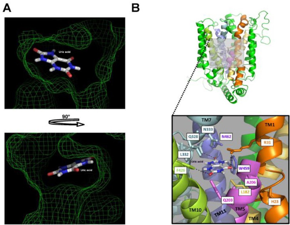
Fig. 5. The uric acid binding site of hGLUT9a. A: The surface was modeled at 3Å around the uric acid substrate forming a binding site pocket (green mesh). B: The putative binding site is formed by amino acids: H23, R31, L182, Q203, A206, Q328, L332, N333, F426, W459 and N462 represented with sticks. Potential interactions of uric acid with R31, Q328 and F426 are shown (dotted lines).